

Ultrasonography
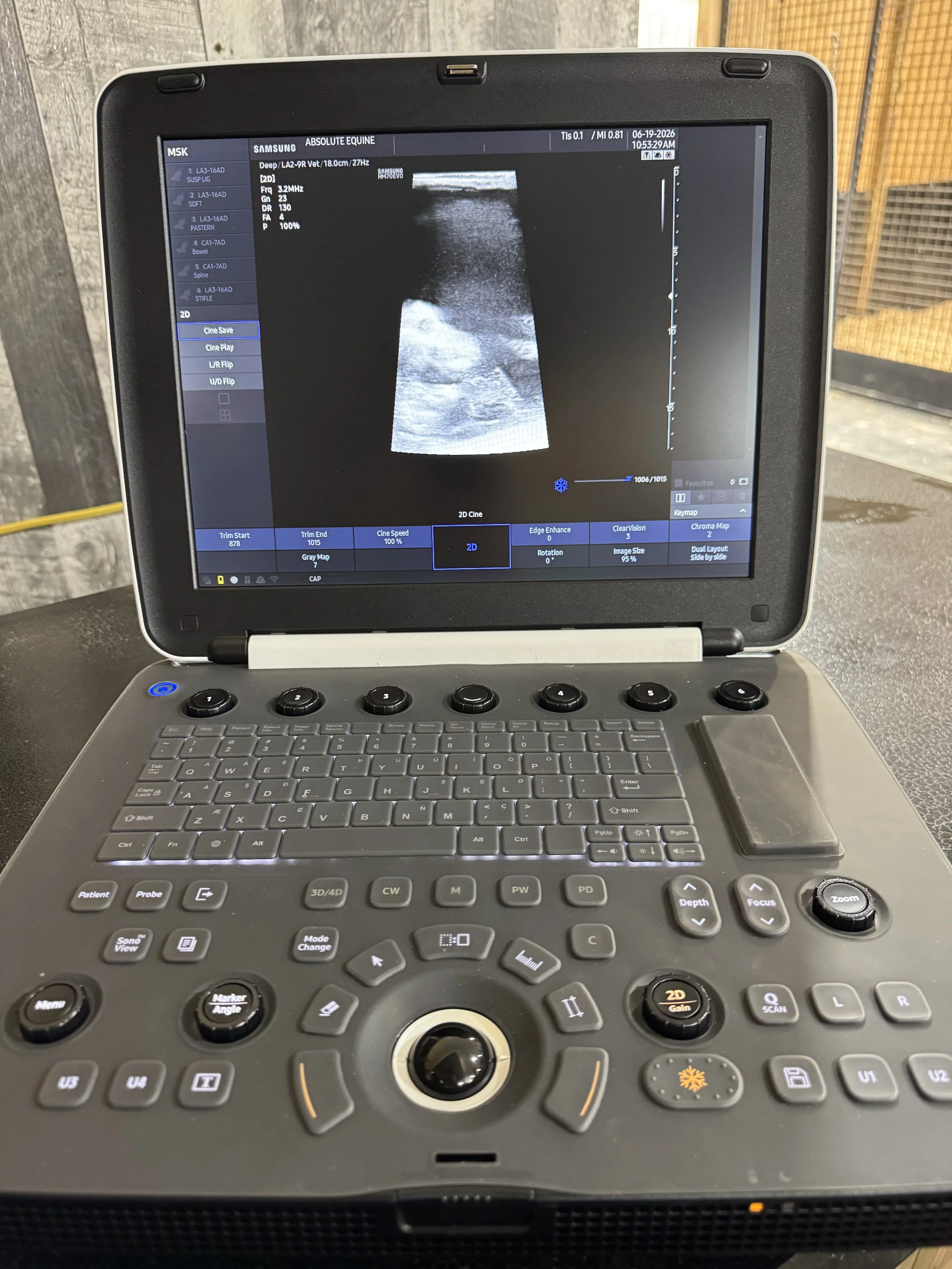
Although radiographs are valuable for evaluating bony structures, ultrasound is probably one of the most versatile tools available in veterinary medicine, and is the diagnostic of choice for evaluating soft tissues. Absolute Equine has a digital ultrasound machine and variety of ultrasound probes available for portable use in an ambulatory setting. We use our ultrasound daily for evaluation of tendons and ligaments, breeding management of mares, pregnancy detection and evaluation, fetal monitoring, measurement of fetal orbit for determination of fetal gestational age, abdominal ultrasonography, and for evaluation of any soft tissue swelling, lump or bump.
Lameness:
We commonly perform ultrasonography of tendons and ligaments in the field setting. We often select the ultrasound after either our physical exam reveals swelling or pain in a tendon or ligament, or after a lameness exam and nerve blocks have helped localize the source of pain to a particular area. Ultrasonography can also be used to evaluate certain synovial structures such as the digital sheath.
Reproduction:
One of the mainstays of reproductive management in the mare is evaluation of the reproductive tract via rectal palpation and ultrasonography. We use it to monitor a mare’s follicular growth, uterine edema, ovulation, and presence of any intra-uterine abnormalities such as endometrial cysts or fluid. With our machine, the equine embryo may be visualized as early as day 12, but we typically suggest the first pregnancy check at day 14 post ovulation. One significant benefit to ultrasound (compared to palpation alone) is the ability to detect and intervene in the case of twin embryos. Some data suggest that 94 percent of twin pregnancies result in abortion or stillbirth of both fetuses. By day 24, the heartbeat can be visualized to verify the presence of a viable fetus.